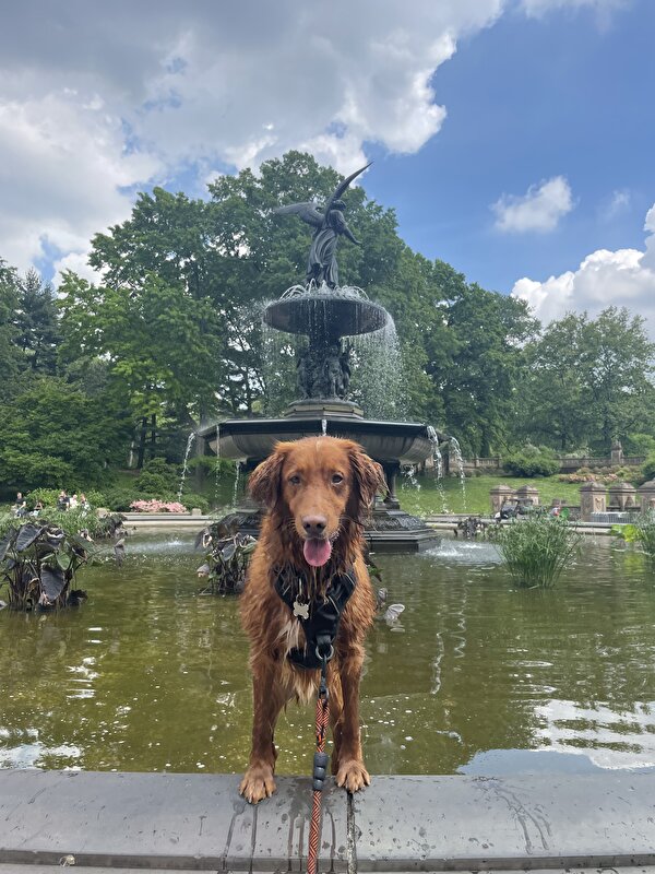
Mijn hond begon 3 weken opeens met hoesten maar voelde zich nog wel goed. Ik dacht dat het kennelhoest was en na 1,5 week leek het beter te gaan totdat hij opeens hoge koorts kreeg, veel slijm aan het ophoesten was, echt ziek was etc. We zijn toen met hem naar de emergency vet geweest en daar hebben ze een scan van zijn longen gemaakt maar deze waren in orde. Hij heeft toen antibiotica meegekregen. Hij werd bij de dierenarts ook steeds slechter, hij kon moeilijk om zijn achterpoten staan. Eenmaal thuis viel hij in diepe slaap en reageerde hij nergens meer op. De volgende dag was zijn koorts gelukkig weg.
Afgelopen donderdag ben ik nog een keer naar de dierenarts geweest want hij was erg benauwd en ademde heel snel (denk door zijn verkoudheid?). Dierenarts gaf aan dat ze geen vermoeden had dat nu een longontsteking was en zijn antibiotica kuur af te maken, hem te stomen in de badkamer etc. Stomen doe ik en helpt hem wel denk ik...
Benauwdheid was volgende dag weer over maar hij is nu na 8 dagen antibiotica (moeten er nog twee) nog steeds erg lusteloos, slaapt veel, veel reverse sneezing, rode ogen, waterige neus, en hoest sinds vandaag weer veel wit slijm op. Zijn eetlust is minder maar met kip eet hij zijn brokjes wel.
Hij is nu dus in totaal al 3 weken niet lekker en lijkt me erg lang voor een kennelhoest. Ik zat ook te denken aan de hondengriep, die geloof ik iets extremer is en hier meer voorkomt (woon in Amerika). Maar ik maak me nog steeds erg zorgen om hem. Ik zou graag de juiste stappen willen nemen mocht hij niet opknappen of wellicht weer zieker worden. Misschien heeft iemand een ander idee wat het zou kunnen zijn? De dierenartsen hier komen met hele lijsten en prijzen aan wat we zouden kunnen doen maar vind het moeilijk om een gerichte aanbeveling te krijgen. Laatste dierenarts beveelt aan, als hij niet opknapt om onderzoek te doen of het kennelhoest, hondengriep etc is, en daarna wellicht een nieuwe scan van zijn longen.
Dierenarts is gewoon ontzettend duur hier (deze twee bezoeken waren $1600!!) maar natuurlijk wil ik er alles aan doen om hem beter te krijgen...
Dit kwam er uit bij zijn bezoek aan de emergency vet een week geleden mocht iemand dat interessant vinden.
Problems List and Differential Diagnosis
1) Fever r/o infection vs inflammation vs stress vs pain vs neoplasia vs immune-mediated vs environmental vs idiopathic vs other
2) Cough r/o: pneumonia vs canine infectious tracheobronchitis (kennel cough) vs foreign body vs bronchitis vs pulmonary edema (cardiogenic/non-cardiogenic) vs parasitic (lung worms; heartworms) vs idiopathic vs neoplasia vs other
3) Lethargy
4) Hyporexia
5) Previous nasal discharge at home
6) Intermittently shaking
7) Leukocytosis: r/o inflammation (infection, immune-mediated disease, tissue necrosis, neoplasia) vs.
corticosteroid-induced neutrophilia/stress leukogram (hyperadrenocorticism/Cushing’s disease, exogenous
steroids, stress) vs. paraneoplastic syndrome vs. epinephrine-induced physiologic leukocytosis vs. chronic or acute leukemia vs. other
8) Neutrophilia: r/o inflammation (infection, immune-mediated disease, tissue necrosis, neoplasia) vs.
corticosteroid-induced neutrophilia/stress leukogram (hyperadrenocorticism/Cushing’s disease, exogenous
steroids, stress) vs. epinephrine-induced physiologic neutrophilia vs. paraneoplastic syndrome vs. granulocytic
leukemia (chronic myeloid leukemia, acute myeloid leukemia) vs. other
9) Monocytosis: r/o inflammation (infection, immune-mediated disease, tissue necrosis, neoplasia) vs.
corticosteroid-induced monocytosis vs. chronic or acute monocytic/myelomonocytic leukemia vs. immune-
mediated neutropenia vs. other
10) Elevated MPV: corticosteroid-induced vs. increased platelet production (ITP vs. in aflammation vs. non-
hemopoeitic neoplasia vs. iron deciency vs. rebound vs. reactive thrombocytosis vs. hemic neoplasia/clonal
thrombocytosis) vs. breed-related macrothrombocytopenia/giant platelet disorder vs. artifact (prolonged exposure >24 hours to EDTA can cause platelet swelling) vs. other
Recommendations
1) Recommended CBC/Chem/Lytes, PCV/TS, BG, lactate, radiographs, respiratory PCR, pulse-ox, hospitalization.
Discussed concern for possible pneumonia or other problems.
-Made owners estimate & discussed with them. O's elect to proceed with CBC & radiographs & go from there.
Progress Notes
1) CBC:
-HCT 50.2%, WBC elevated @ 27.72 (5.05-16.76 K/uL), neutrophils elevated @ 23.98 (2.95-11.64 K/uL), monocytes
elevated @ 1.21 (0.16-1.12 K/uL).
-Lactate: 1.8 mmol/L.
2) Administered 0.3 mg/kg butorphanol IV for light sedation for radiographs & 1 mg/kg Cerenia IV.
3) Radiographs:
Conclusions:
Unremarkable thorax.
Mild prostatomegaly: The primary differential is benign prostatic hyperplasia, not unexpected for the signalment.
Active prostatic disease (prostatitis, cystic infiltrates) are possible. Correlate with rectal exam findings.
Postprandial but otherwise unremarkable abdomen.
Comments:
The cause of the respiratory signs is not clearly apparent from this study. No active bronchopulmonary inflltrate is seen, but this does not exclude underlying inflammatory disease. Differentials include small airway disease (sterile chronic bronchitis), environmental irritants, infectious tracheobronchitis/CIRD, parasitic bronchitis, eosinophilic bronchopneumopathy, all of which may present with minimal radiographic lung changes. If the cough persists or progresses in the absence of systemic signs then an empirical treatment trial for infectious bronchitis could be considered. A canine infectious respiratory disease PCR panel could also be considered.
Alternatively, airway washing or brushing for cytology and culture would be helpful in determining the nature of any underlying active bronchopulmonary infiltrate.
Follow-up thoracic radiographs may be indicated if respiratory signs progress.
The cause of pyrexia and leukocytosis is also not clear, and may be unrelated to the more chronic cough.
In addition to the hematology and if not already performed, baseline biochemistry and urinalysis could be
considered. The breed is also predisposed to hypoadrenocorticism, but this would be an unusual presentation.
An upper respiratory tract component is also possible: Direct imaging of this region may be helpful.
If the pyrexia persists, full abdominal ultrasound may be helpful in screening for an intra-abdominal in inflammatory focus.
3) Pulse-ox on room air: 100%
4) Called O's to discuss all results ~14:10.
-Discussed hospitalization for Bowie (IV fluids, IV antibiotics, sometimes need oxygen therapy, etc)., possible
further diagnostics. Owners elect to take him home to monitor at home. Told owners multiple times if not
improving or worsening (ex: lethargy, anorexia, tachypnea and/or dyspnea, etc.) that hospitalization is recommended +/- further diagnostics may be recommended (ex: respiratory PCR, possible airway sampling @ specialty and/or transfer to specialty hospital, etc.). Discussed with owners that if not eating, cannot receive antibiotics; hospitalization is again recommended. O's understood. Discussed that sometime, radiographs can lag behind clinical signs.
-Rectal examination: No significant findings. Prostate palpates normally.
-O noted shaking of hind limbs & sinking in the hind end when standing and asked if it is a side effect of the
medication given. Discussed it is possible, or can be nervous, uncomfortable, etc. O's to monitor at home.
-Administered 650 mLs LRS SQ.
-Medications to go home: Clavamox, ondansetron.
een foto van hem in betere tijden
