Anoniem schreef:verootjoo schreef:Zijn er echt nog mensen die denken dat autisme met vaccinaties te maken heeft?
Ik heb daar nog geen uitsluitsel over. Heb dit laatst gevonden en wil het nog allemaal lezen.
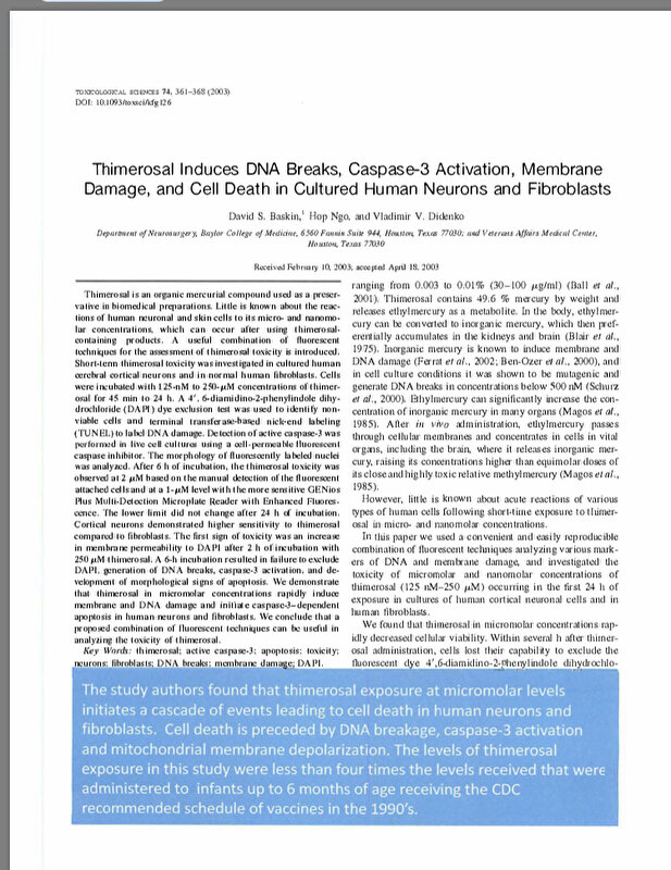
https://childrenshealthdefense.org/wp-c ... 4.2.18.pdf
(In het losse tetanus vaccin zit thiomersal, daarom heb ik laatst ervoor gekozen alleen de tetanus TIG te doen.)
https://www.google.nl/amp/s/amp.nos.nl/ ... tisme.html
En dit is er nog maar eentje. Hoe kán je dit nog geloven. Als je even door de rest van de site heen kijkt valt hij voor mij af als valide bron. De angstzaaiende koppen, het feit dat het een particiliere website is en het team geen enkel ook maar een beetje medisch gekwalificeerd persoon heeft (ze zijn vooral goed in marketing)... ik vind het moeilijk te begrijpen dat je de info op een site als dit wel gelooft maar biv. Het RIVM niet.
En het gaat niet over identificeren met wat je schrijft, het maakt wel uit. Je gooit bangmakende onwaarheden uit onderzoeken die je (zo blijkt) verkeerd interpreteerd de wereld in. Dat je zelf niet vaccineert op medische gronden, helemaal prima. Doordat we een hoge vaccinatiegraad hebben is dat geen enkel probleem. Maar jouw situatie is een uitzondering en wil niet zeggen dat vaccinaties gevaarlijk zijn, alleen dat ze op jou een slecht effect hadden (en zelfs dat weet je ook niet zeker).